1. Sample collection
Collect the infected fruits, leaves etc. from the horticulture garden. Put them in the food preservation bags with a piece of wet paper towel to keep the humidity.

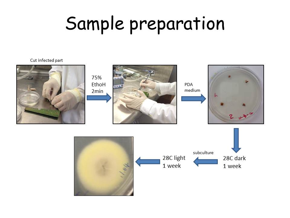
2. Sample preparation:
Cut the infected part of samples into several small pieces and sterilize surface in 75% EthoH for 2 min.
Rinse the samples in autoclaved water, and then put on the PDA medium.
Put under 28C incubator to grow in dark for 1 week, and then transfered under light to induce the sporulation.
Check the unknown fungi structure under microscope when they sporulate.
3. Unknown fungi 1

The unknown fungi #1 is isolated from the infected pepper.
There is no beautiful hyphae grown on the PDA medium. I just mount a sample from a black spot. Check the squash mount under the microscope.
At first, I thought this is just a common fungi, Fusarium. However, the spores do not have the septa, which is different that of Fusarium.
When I see through the whole sample, Dr. Shaw and Dr. Ebbole helped me to recognize this Setae structure, which is a obvious structure for Colletotrichum. Then I googled and found Colletotrichum capsici can infect pepper. Hence, this isolate is diagnosed as Colletotrichu capsici.

Colletotrichum can cause anthracnose on pepper, grapes etc., causing small areas of dead tissue on the host. It is a popular pathogen cause economical damaged disease, anthracnose.
4. Unknown fungi 2

The unknown fungi #2 is isolated from one contaminated PDA plate. The structure of this fungi is so distinctive. There are cylindrical vesicles forming at the apex of the cob-like sporangiophores contained sporangia.

By comparing with the fungi key book, it is diagnosed as Mycotypha indica, which belongs to Zygomycota.
5. Unknown fungi 3


This is a relative common filamentous fungus, diagnosed as Curvularia sp., which has been identified by many people in our class. It is a facultative pathogens from the ascomycota.
6. Discussion:
At first, I thought the unknown fungi isolation project must be a hard-to-achieve mission. I didn't expect that in the end I could identify several species and pick 3 not from the "Group 1". I am grateful to many Dr. Shaw and Dr. Ebbole. Also, Xin, Wenwei and David helped me a lot during the process. To me, this is a real case of "nothing is impossible".
hi... can u share with me your protocol to mount the fungal colony onto the slide for microscopic viewing?
ReplyDeletejust use an autoclaved needle to pick a very tiny amount of the fungi. put it on a glass slide with a drop of water . then place a cover slide on it.
DeleteSorry for replying late.
Hi... I have a few mold image under microscope need to be identified. can u help me to identify them? or share with me how u identify them? let say, what kind of resources u using? or journals? or website?
Deleteor can i send the picture to u, we discuss about it together...