1. Using the fluorescence microscopy to check protein localization
2. Observe fungal structure (Buller's drop and clamp connection)
1.1 The GFP tagged protein expressed in nucleus
 |
| GFP tagged protein expressed in nucleus |
Images captured from the video recorded under fluorescence microscopy. Since the video doesn't work, I use the pictures instead.
 |
| Elapse picture for hyphal growth Spitzenkorper can be visualized as a brighter spot at the hyphal tip |

GFP-tagged protein under fluorescence microscope
The GFP-tagged protein moving through septum.
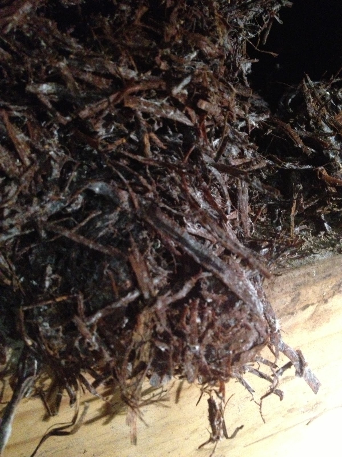
2. Observe the micro structure of Chlorophyllum sp.
 |
| Chlorophyllum sp. |
 |
| Basidiospores |
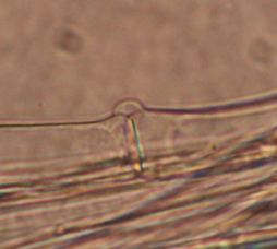 |
| Clamp connection |
Clamp connection is unique to Basidiomycota to ensure each cell is binucleate, but not all the basidiomycete have this phenomenon. It happens in the dikaryons stages formed by the terminal hyphae.

a&b The clamp connection is formed in the terminal hyphae when the hyphae is long enough.
c The 2 nuclei undergo a mitotic division.
d The b nucleus move to the new clamp and a septum forms to trap nucleus b. The a' and b' nucleus migrate to the hyphal tip, while the a nucleus migrate away from the hyphal tip.
e Then a septum below the clamp connection formed to form a new cell containing nuclei a' and b'. The clamp fused to the old cell release b nucleus.
In the end, there are 2 compatible nuclei in each cell to ensure each cell is binucleate.
http://www.botany.hawaii.edu/faculty/wong/Bot201/Basidiomycota/Clamp_connection_formation.htm
Buller's drop:
Though we are not able to observe the buller's drop. It is very worthwhile for us to study this interesting spore releasing mechanism. This spore dispersal mechanism is determined by Dr. A. H. R. Buller.
I cited the picture to illustrate Buller's drop as follows:

Pic. a A buller's drop, a fluid drop, is formed at the base of the spore by the condensation of water resulting from spore's low water potential caused by high sugar concentrtion.
Pic. b The ballistospores will be discharged from mushroom gills in the predicted route carried by the buller's drop.
Pic. c Successive images of how the buller's drop helps release the ballistospores.