Friday, November 30, 2012

20121024 Fluorescence microscopy usage and Fungal structure observation

Goals:
1. Using the fluorescence microscopy to check protein localization
2. Observe fungal structure (Buller's drop and clamp connection)


1. Usage of the fluorescence microscopy
1.1 The GFP tagged protein expressed in nucleus

GFP tagged protein expressed in nucleus
1.2 Elapse images
Images captured from the video recorded under fluorescence microscopy. Since the video doesn't work, I use the pictures instead.

Elapse picture for hyphal growth
Spitzenkorper can be visualized as a brighter spot at the hyphal tip

 GFP-tagged protein under fluorescence microscope 
The GFP-tagged protein moving through septum.


2. Observe the micro structure of Chlorophyllum sp. 
Chlorophyllum sp. 

Basidiospores
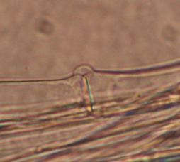
Clamp connection:
Clamp connection

Clamp connection is unique to Basidiomycota to ensure each cell is binucleate, but not all the basidiomycete have this phenomenon. It happens in the dikaryons stages formed by the terminal hyphae.  

a&b The clamp connection is formed in the terminal hyphae when the hyphae is long enough. 
c The 2 nuclei undergo a mitotic division. 
d The b nucleus move to the new clamp and a septum forms to trap nucleus b. The  a' and b' nucleus migrate to the hyphal tip, while the a nucleus migrate away from the hyphal tip. 
e Then a septum below the clamp connection formed to form a new cell containing nuclei a' and b'. The clamp fused to the old cell release b nucleus. 
In the end, there are 2 compatible nuclei in each cell to ensure each cell is binucleate.

http://www.botany.hawaii.edu/faculty/wong/Bot201/Basidiomycota/Clamp_connection_formation.htm


Buller's drop:
Though we are not able to observe the buller's drop. It is very worthwhile for us to study this interesting spore releasing mechanism. This spore dispersal mechanism is determined by Dr. A. H. R. Buller.
I cited the picture to illustrate Buller's drop as follows:


Pic. a A buller's drop, a fluid drop, is formed at the base of the spore by the condensation of water resulting from spore's low water potential caused by high sugar concentrtion.
Pic. b The ballistospores will be discharged from mushroom gills in the predicted route carried by the buller's drop.
Pic. c Successive images of how the buller's drop helps release the ballistospores.

http://openi.nlm.nih.gov/detailedresult.php?img=2612744_pone.0004163.g001&query=the&fields=all&favor=none&it=none&sub=none&sp=none&req=4&simCollection=3092991_hir-17-29-g001&npos=45&prt=3

20121017 Lab 7 Continuation of N. crassa genetics

Goals:
1. Continuation of Neurospora genetics 
2. Observation of field collected fungal samples


1. Continuation of Neurospora genetics 
Steps:
1.1. Take out the Neurospora plate where 2 strains crossed.
Successful cross for Neurospora Crassa

Neurospora crassa under dissecting microscope
 1.2. Pick the perithecium by using a dissecting needle put on glass slide with a drop of autoclaved water and then made a crush mount. Observe under the microscope


2. Observation of field collected fungal samples


Morel: 
Morchella genus, Pezizomycetes class, Ascomycota. 
The honey-comb shaped ascocarps are prized by french cuisine. The Morchella has the antioxidant function and can improve the liver function. Also, it is reported that it contain high macronutrient level, like fiber, which provides cardiovascular and weight loss support. Besides, it also contains high vitamin level.

http://www.livestrong.com/article/446035-what-are-the-health-benefits-of-morel-mushrooms/


Stinkhorn mushroom:
Phallaceae, Agaricomycetes class, Basidiomycota
Stinkhorn mushroom is believed to be a smelly but beneficial fungi. It produces a foul, meat-rotting smell to attract the insects to help them with spore spreading. Though, the smell may be putrid, it indeed help the garden with breaking down the mulch and enable the nutrient to be absorbed by the plants.

Reference from wikipedia

Bird's nest fungi:
Nidulariaceae family, Agaricomycotina subphylum, Basidiomycota
The Bird's nest fungi usually resemble the bird's egg-filled-in nest. It is saprobic and usually can be found on the decay wood. The bird's nest fungi are famous for the special mechanism of spore dispersal. The egg shaped structure contains the spores, which are called as peridioles. Dr. Harold Brodie determined the mechanism as force can released the spores and the spores can attach to a substrate, which enable the spores eaten by an animal and transported to everywhere.


Reference from wikipedia

http://www.botany.hawaii.edu/faculty/wong/BOT135/Lect05_c.htm (interesting website describing all kinds of mechanisms of spore dispersal in fungi)

Isolation of Unknown Fungi

1. Sample collection

Collect the infected fruits, leaves etc. from the horticulture garden. Put them in the food preservation bags with a piece of wet paper towel to keep the humidity.
2. Sample preparation:
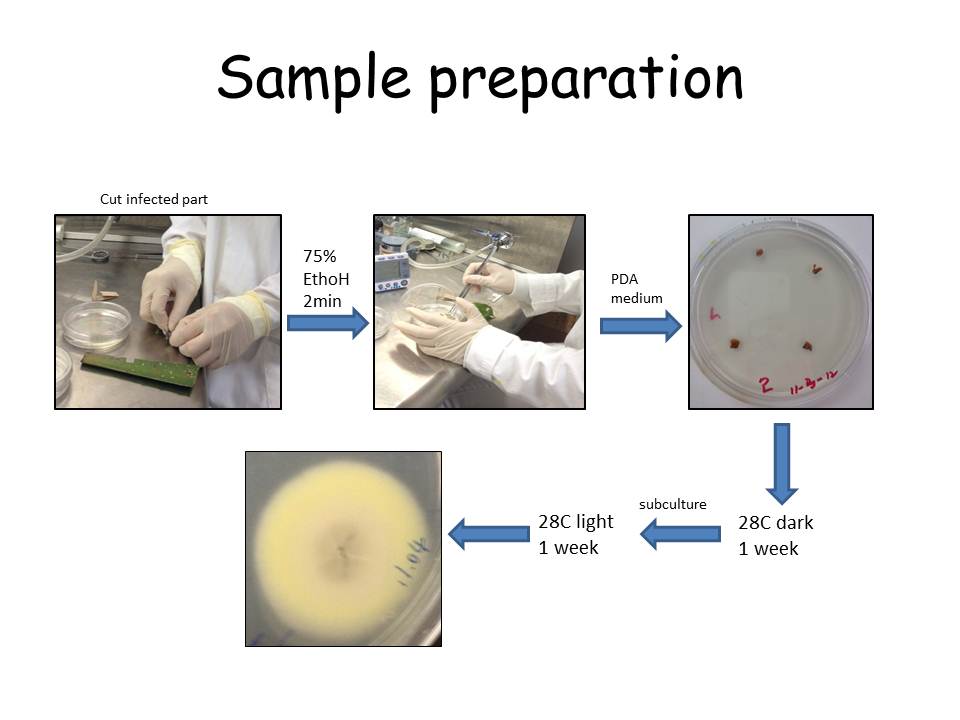
Cut the infected part of samples into several small pieces and sterilize surface in 75% EthoH for 2 min.
Rinse the samples in autoclaved water, and then put on the PDA medium.
Put under 28C incubator to grow in dark for 1 week, and then transfered under light to induce the sporulation. 
Check the unknown fungi structure under microscope when they sporulate.
3. Unknown fungi 1


The unknown fungi #1 is isolated from the infected pepper. 
There is no beautiful hyphae grown on the PDA medium. I just mount a sample from a black spot. Check the squash mount under the microscope. 
At first, I thought this is just a common fungi, Fusarium. However, the spores do not have the septa, which is different that of Fusarium
When I see through the whole sample, Dr. Shaw and Dr. Ebbole helped me to recognize this Setae structure, which is a obvious structure for Colletotrichum. Then I googled and found Colletotrichum capsici can infect pepper. Hence, this isolate is diagnosed as Colletotrichu capsici


Colletotrichum can cause anthracnose on pepper, grapes etc., causing small areas of dead tissue on the host. It is a popular pathogen cause economical damaged disease, anthracnose. 


4. Unknown fungi 2


The unknown fungi #2 is isolated from one contaminated PDA plate. The structure of this fungi is so distinctive. There are cylindrical vesicles forming at the apex of the cob-like sporangiophores contained sporangia. 


By comparing with the fungi key book, it is diagnosed as Mycotypha indica, which belongs to Zygomycota. 

5. Unknown fungi 3

The unknown fungi #3 is isolated from tree leaves. There are pigmented spores forming on the geniculate conidiophores in a sympodial manner.
This is a relative common filamentous fungus, diagnosed as Curvularia sp., which has been identified by many people in our class. It is a facultative pathogens from the ascomycota.

6. Discussion:
At first, I thought the unknown fungi isolation project must be a hard-to-achieve mission. I didn't expect that in the end I could identify several species and pick 3 not from the "Group 1". I am grateful to many Dr. Shaw and Dr. Ebbole. Also, Xin, Wenwei and David helped me a lot during the process. To me, this is a real case of "nothing is impossible".

Monterey Mushroom Facility Tour

What is Monterey?

I bet you must see it in the supermarket!
Oyster Mushrooms Shiitake Mushrooms

Today, our course is to visit this big mushroom factory. Cool!


For food safety issue, everyone is required to wear a hair net, toes covered shoes, no shorts,bno jewelry, no stuff in the pocket.

1. This machine transport the compost the straw mixture with the chicken poop and urea from outside into the mushroom growth chamber.


2. The environmental factors like temperature, light, humidity are monitored by the computer, but also needs people to check several times every day. 


 3. A pile of wooden trays containing compost inoculated with mushroom mycelium in the growth room with optimal temperature and high humidity.

Wooden trays contained sanitized compost

Packaged millet seeds pre-inoculated with mycelium

Compost with inoculated mycelium

Mushroom begins to grow on the nutritious compost

4. When the mushroom grow to certain size, they will be transported to other growth room set with the optimal environmental factors. The instructor said they will transport the whole pile of wooden tray at one time. How do they make it? I am really curious about this.

5. At last, they are transported into the growth room waiting for harvesting. 

Clean environmental

What a lovely mushroom!

The worker is selecting the mushroom with high quality and 
separate then into different colored plastic basket, indicating the different quality levels.
They indeed discard the mushroom with bad quality!!


6. The environment needs to be very clean, but fungicide is the last thing used to kill pathogens.
If there is contamination in the compost, the workers will use salt to cover the contaminated area.


7. At last, I know how do they transport the whole pile of wooden tray at one time!! They use this machine (sorry! I forgot the name...). Lorna told me that the drivers need special driver license to operate on this machine for this may hurt people because of wrong operations. Wow! He is very proficient in operating the machine! It is really cool!!


Discussion: 
Monterey, established in 1971, is the country's largest maker of mushrooms. It is headquartered in Watsonville, California. They are selling all kinds of mushrooms. After this tour, I feel safer to eat there mushroom. 

White Mushrooms  Portabella Mushrooms
Organic Mushrooms Specialty Mushrooms

Without this great opportunity, I bet I will never imagine how does the industry produce the mushroom! Thanks to the lab course. Hope it will be custom for this class for the students love it so much!


Oh, my God! It is so fresh!! 
I promise this is the first time for me to each raw mushroom!!


Fruit mushroom by using the mushroom kits


1. Goal:
Fruit mushroom by using the Gourmet Mushroom Kits for the Home.

2. Materials:
The Pearl Oyster Mushroom Patches
The Shiitake Mushroom Patch
Sticks
tapes
Water spray bottle

3. Introduction:
The Pleurotus ostreatus (Oryster mushroom) belongs to the Pleurotus genus, Pleurtaceae family, Agaricales order, Agaricomycetes class, Basidiomycota phylum.
They are gilled mushroom and have the clamp connections. Also, they can be used in mycoremediation in petroleum. It is white-beige form, grown between 55-75 F, which thrives on poplar and willow.

The Lentinula edodes (the Shiitake mushroom) belongs to the Lentinula Genus, Marasmiaceae family, Agaricales order, Agaricomycetes Class, Basidiomycota phylum.
Consumption of raw or slightly cooked shiitake mushrooms can cause Shiitake dermatitis, caused by the toxin lentinan.


Oryster mushroom


    Shiitake mushroom
 4. Instructions:
4.1 Oryster mushroom
Step 1.Open the kit and read the instructions. 
Surprisingly, we found that there is there are some octopus/coral form oyster mushroom in the kit box when we open it. These octopus form of oyster mushroom is caused by the light starvation and they are edible when fresh.


Step 2. Tie the sticks to form a steady frame to support the plastic bag called humidity tent


Step 3. spray some water on the mushroom patches to keep it moist.

 Step 4. Pull the humidity tent over the frame to keep the mushroom patches moist.
Step 5. Put the whole mushroom patch under room temperature on the top of the microscope case nearby the window and spray with water 3 to 5 times a day.

4.2 Shiitake mushroom

When we open the kit, the Shittake mushroom looks like popcorn. Hence, according to the instructions, we soaked the shiitake mushroom  in water for 3 hours. Then we cover it with the humidity tent. Put it by the window but not exposed to direct light. Spray with water for 3 to 5 times per day. 


5. Observation: 
1 week later, we observed that there are several mushroom come out from the mushroom patches.

Oryster mushroom

Shiitake mushroom

 6. Discussion:
6.1 Moisture
We keep moisture, the key factor for fruiting the mushroom, by spraying it w water 3-5 times a day. The water can be from spring or boiled tap water. Chlorinated or distilled water is not recommended. Chlorinated water will discourage the growth of mushrooms, while the distilled water lacks nutrient.

It is written in the instructions as that "The more you mist the mushroom patch, maintaining a high humidity zone around it, the more bountiful your crops will be." 

6.2 Light:
All oyster mushrooms need light to grow but the ideal place to produce is not exposed to direct sunlight.