Wednesday, October 10, 2012

Lab 2 20120919

1.  Aspergillus
We learned the difference between Bisereate and Unisereate that the Bisereate has metulae on the vesicles.


1.1. Aspergillus nidulans

Pic 1 Aspergillus nidulans (bisecreate)

Pic 2 Aspergillus nidulans (unisecreate)

1.2. Aspergillus tamari

Pic 3 Aspergillus tamari (bisecreate)

Pic 4 Aspergillus tamari (unisecreate)

1.3. Aspergillus paraciticus
Pic 5 Aspergillus paraciticus

1.4. Aspergillus sojae
Pic 6 Aspergillus sojae

1.5. Aspergillus flavus
Pic 7 Aspergillus flavus

1.6. Aspergillus oryzae
Pic 8 Aspergillus oryzae


2. Mediums
2.1 1/2 PDA
Potato dextrose agar
Almost everything can grow on it.

2.2 Water agar
Only agar with low nutrient. Some fungi sporulate better under low nutritious condition. They may try to get rid of the bad conditions.

2.3 Corn meal
It is good for plant pathogen to grow.

2.4 Rose Bergal
It has red color and have to be stored in dark, containing some antibiotics.
It is good for samples collected from the soil.

Lab 3 20120918


1. Riddell Mount:
Last week, we learned an important mount method: riddell mount.
Microscopic preparations are essential for an accurate identification of an organism. However, fungi that have delicate sporing structures are easily disturbed upon removal.
Compared to squash mount and tape mount, slide culture is a better method, which involves growing the fungus on a plug of agar, which is resting on a coverslip; the coverslip can then be mounted directly onto a microscope slide.
The obvious drawback of this method is the short life span of the fungus because of the limited nutrient source.


 Pic 1 Riddell Mount of Aspergillus flavus

Pic 2 Riddell Mount of Aspergillus niger

2. Phytophtrora infesta 
2.1 Phytophtrora infesta sporangiaphore

Pic 3 Phytophtrora infesta sporangiaphore
2.2 Phytophtrora infesta zoospore


Video Phytophtrora infesta zoospore

From the video, we can see the movement of zoospores. However, I didn't see the clearly about the mophology of the zoospores.

This mount got contamination unfortunately....

Lab 1 20120905


1. Tips for using microscope
Keep in mind that whenever we should transport the microscope gently with 2 hands holding it and use the body to support it too.

2. Mounts type:
Squash mounts: Add a drop of water on the glasses. Use sterilized scapel to cut a small piece of agar with colonies on it. Put a cover slide and then squash the agar gently. It is easier to observe the spores since the outside force push the fungi release spores.
Tape mounts: when we want to see the intact fungal structure, like sporophore, it is better to use tape mount by just using the tape contact with the hyphal structure on the plate gently. Then, put the piece of tape on the glass slide with a drop a water on it. By using tape mounts, you can save the intact structue but we have to handle it really gently.

Other ways:
We can even put the agar directly on the slides.
We can also use the innoculating needle to pick a small amount of colonies and suspend in the water, then cover with cover slide, but do not let the cover slide sit on the colonies directly by putting small pieces of glasses on the glass to support the cover slide in order to leave some space for the fungi and keep the intact structure.

3. Have a general review of fungal world
3.1 Alternaria brassicicola
 Pic 1 Aspergillus conidia chain


Pic 2 Aspergillus conidia

3.2 Cladospsorium Sp. 
Pic 3 Cladospsorium Sp. (Upper: squash mount, Lower: tape mount)

With squash mount, I can see the hyphal fragment.
With the tape mount, I can see the intact structure of Cladospsorium Sp. conidiaphore.


Pic 4 Cladospsorium Sp. conidiaphore

3.3 Thielaviopsis bassiola 
Thielaviopsis bassiola has 2 types of asexual spores: phialospore (endospores) and aleuriospore(chlamydospores). Chlamydospores are produced in chains (two to eight spores per chain). The dark, thick walled chlamydospores separate at maturity and each cell is capable of germination.
Endoconidia are produced in greater numbers than the chlamydospores. These spores are variable in size with slightly rounded ends. The septae hyphae are hyaline at first and become pigmented with age.  A sexual state has not been observed with T. basicola. 

Pic 5 Thkelaviopsis bassicola conidiaphore

3.4 Pythium ultimum (oomycete)
 Pic 6 Pythium ultimum

Pic 7 Oogonium (female)

The oogonium after fertilization will turn into oospore (thick wall)

4. Hemocytometer
We can first dilute the spore suspension to a certain concentration and then use trypan blue to stain the fungi and then count on Hemocytometer under microscope.

Lab is about having fun!


See how professional we are!
Sorry for the picture of Danny~~
Sorry for Sheila was not there that day~
Anybody wants his or her picture, just let me know your email address.

Have fun!


Lab 4 20120926


1. Neurospora crassa (Ascomycete)
Goals: Observe ascospores, microconidia (only produced on poor nutritious agar plate) and macroconidia.
Neurospora crassa is a type of red bread mould and the best-characterized filamentous fungi. It has a relatively simple haploid life cycle. It contributes to biological research by being a model system. It helps Dr. Beadle and Dr.Tatum to raise the "one gene, one enzyme" hypothesis and have been a very useful tool for people to study Genome evolution, circadian rhythm, epigenetics etc.
During the asexual stage of Neurospora crassa, they produce both microconidia and macroconidia. Meanwhile, they produce ascospore as the sexual spores, which unfortunately failed to found by me in the class.

1.1 Macroconidia chain

 Pic 1. Neurospora crassa Macroconidia chain
I can see the intact macroconidia chain by using tape mounts.

1.2 Hyphae

Pic2. Neurospora crassa Hyphae

1.3 Microconidia

Pic 3 Neurospora crassa microconidia (small spores circled with red lines)
It is much easier to find macroconidia than microconidia. The microconidia can be only produced on poor nutritious agar plate.

2. Neurospora crassa mating
For heterthallic Neurospora crassa, they need two mating type spores come into contact, MATA and Mata. In our lab practice, we have smRP10 as MATA strain and smRP11 and NCAC021-1 as Mata strains.

Pic 4 Preparation for mating

2.1 Prepare the strains and medium plates used for mating, and divided the medium plate into half with a Sharpie pen.
2.2 Handle the mating process by the burner and sterilize all the tools like the inoculating needle.
2.3 I picked a little bit spores of SMRP10 and inoculate on one side of the medium plate and then steriled the inoculating needle and picked small amount of spores of SMRP11.
2.4 Sealed with the parafilm, and then put the plate under 25C.
2.5 I also did a control by mating SMRP11 and NCAC021-1, which are 2 MATa strains, and supposed to failure of mating.
2.6 One week later, we observed the mating results. We can see successful mating between SMRP10 and SMRP11, which belong to 2 different mating types. The black dots in the picture below indicates sucess of mating, while no mating product can be observed form the control group. However, the 2 strains didn't mate in the middle of the medium plate perfectly. It may due to spores of one of the strain move faster than the other one.


Pic 5 SMRP10 (A) X SMRP11 (a) successful mating


Pic 6 SMRP11 (a) X NCAC021-1 (a) unsuccessful mating


Pic 7 Perithecia of N. Crassa 
I also observed the black mating product under the dissecting scope.

3. Ustilago (Basidiomycete)
3.1 Observe disease symptom
Pic 8 Disease symptom of Ustilago 1 week after inoculation

3.2 Inoculate the healthy Maize leaves with Ustilago
First, I picked 2 uniform and healthy Maize plants. Then, I used the syringae to inoculate the Maize stem until we can see liquid drop appear around the whirl of the plant. Meanwhile, inoculate another healthy plant with only water.
Pic 9 Healthy plants before inoculation (Left: water, Right: Ustilago)
Pic 10 10 days post inoculation (Left: water, Right: Ustilago)
1 week later, I can see obvious Ustilago tumors on Maize inoculated with Ustilago, while the control plants which only be inoculated with water are still very healthy and green.

3. Mucor Dimorphism (Zygomycete)
When deprived of oxygen, Mucor grows as spherical, multipolar budding yeasts. In the presence of oxygen, they propagate as branching coenocytic hyphae.
Pic 11 Mucor Dimorphism

Lab 5 20121003


1 Stain the Maize leaves with developed tumor symptome due to the inoculation of Ustilago
Cut the Maize leaves showing disease symptom into several piecies, distained in the Ethoh: Acetic Acid=2:1 solution overnight. Then poured out the distaining solution and stained the leaves in the Trypan Blue Dye in lactophenol for 6 hr, then distained the leaves for 1 hr. Check the sample under the compound microscope.
Trypan Blue staining Maize leaves

The Trypan Blue can stain both dead cells and fungal structures. Under the microscope we can see there are indeed existence of Ustilago in the tumor of the Maize leaves.

2 Observe all kinds of spores in Ascomycota
2.1 Tricoderma viride (Ascomycota/Sordariomycetes/Hypocreomycetidae)
Trichoderma viride is a fungus and a biofungicide. It is used for seed and soil treatment for suppression of various diseases caused by fungal pathogens. It is also a pathogen causing green mould rot of onion.

 Pic 1. Tricoderma viride Chlamydospore
The bigger spore I saw should be Chlamydospore, which is a kind of resting cell, more resistant to adverse environment.

 
Pic 2. Tricoderma viride conidia
The smaller spore I saw should be conidia, small, round.

2.2 Pestalotia Sp. (Ascomycota/Sordariomycetes/Xylariomycetidae)
Pestalotia is primarily a secondary pathogen. It is saprophytic on dead and dying tissues and is weakly parasitic infecting wounds under moist conditions, causing important diseases- Pestalotiopsis tip blight of conifers.


Pic 3. disease syptome of tip blight of conifers (from website)

 
Pic 4. Petalotia. sp. conidia

Conidia are multi-celled with usually three darkly pigmented center cells and clear pointed end cells. Conidia are ellipsoid or fusoid (football-shaped). A diagnostic feature is the two or more clear, whisker-like appendages arising from the end cell.
http://plantpath.caes.uga.edu/extension/Fungi/pestalotia.html

2.3 Fusarium graminearum (Dikarya/Ascomycota/Pezizomycotina/Sordariomycetes/Hypocreales)
 The name of Fusarium comes from Latin fusus, meaning a spindle. Fusarium is a large genus of filamentous fungi widely distributed in soil and in association with plants. Most species are harmless saprobes, and are relatively abundant members of the soil microbial community.
Some species produce mycotoxins in cereal crops that can affect human and animal health if they enter the food chain. The main toxins produced by these Fusarium species are fumonisins and trichothecenes. F. graminearum can also cause root rot and seedling blight. The total losses in the US of barley and wheat crops between 1991 and 1996 have been estimated at $3 billion.


 Pic 5. Fusarium graminearum macroconidia

2.4 Colletotrichum coccodes (Ascomycota/Pezizomycotina/Sordariomycetes/Glomerellales)
Colletotrichum coccodes is a plant pathogen, which causes anthracnose on tomato and black dot disease of potato. Many of the species in this genus are plant pathogens, although some species may express a mutualistic life-style in non-disease hosts as obligate symbionts to plants in the form of endophytes.

Pic 6. Colletotrichum coccodes conidia
Colletotrichum coccodes conidia are straight, fusiform, attenuated at the ends.


Pic 7. Colletotrichum coccodes sclerotia with setae
Sclerotia are usually abundant, setose, spherical and are often confluent.
http://www.mycology.adelaide.edu.au/Fungal_Descriptions/Coelomycetes/Colletotrichum/

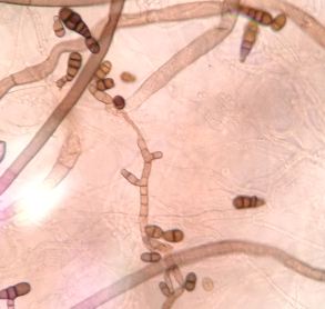
5. Alternaria brassicicola (Ascomycota/Dothideomycetes/Pleosporales)

Pic 8. Alternaria brassicicola 

Alternaria brassicicola is considered a necrotrophic plant pathogenic fungus and like other Alternaria species has been shown to secrete numerous toxic secondary metabolites and proteins that cause cell death via induction of apoptosis in plants or by directly damaging cells. A. brassicicola causes black spot disease on virtually every important cultivated Brassica species including broccoli, cabbage, canola, and mustard.
Alternaria brassicicola has routinely been used as a model necrotrophic fungal pathogen in studies with Arabidopsis thaliana, also a weedy member of the Brassicaceae plant family.
From a human health perspective, Alternaria brassicicola is representative of a genus of fungi that is considered one of the most potent sources of mold-derived allergens. Moreover, sensitization to Alternaria is also strongly associated with chronic respiratory diseases such as asthma and chronic rhinosinusitis.
http://genomeportal.jgi-psf.org/Altbr1/Altbr1.home.html

6. Botrytis Cinera (Ascomycota/Pezizomycotina/Leoitomycetes/Helotiales)
Botrytis cinerea is a necrotrophic fungus that affects many plant species, although its most notable hosts may be wine grapes.
Botrytis cinerea is characterized by abundant hyaline conida (asexual spores) borne on grey, branching tree-like conidiophores. The fungus also produces highly resistant sclerotia as survival structures in older cultures. It overwinters as sclerotia or intact mycelia, both of which germinate in spring to produce conidiophores. The conidia are dispersed by wind and rain-water and cause new infections.


Pic 9. Botrytis cinera